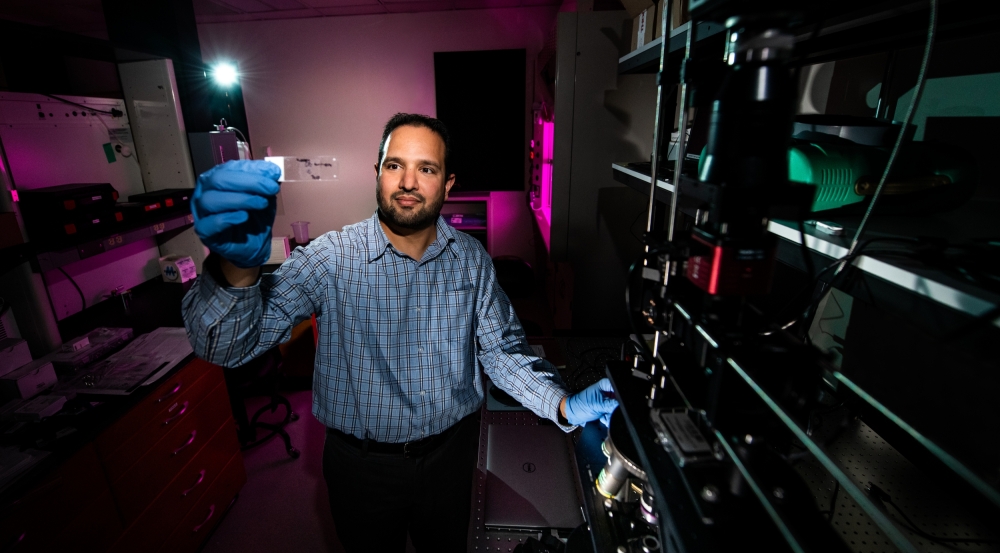
Biophotonic, Electric, Acoustic, and Magnetic Measurement Laboratory

The Biophotonic, Electric, Acoustic, and Magnetic Measurement (BEAMM) Laboratory is a space for experimenting at the interface of technology and biological materials. Staff across divisions at the Laboratory use the lab to support research programs, from scanning cells with a novel hyperspectral microscope to see inside without destroying the cells to using deep learning to analyze images of tissue. The 320-square-foot lab is a Biosafety Level 2 facility, which can support the study of human tissue and cell lines.
In the lab, staff can use a range of technologies and techniques, such as optics, lasers, radio frequency signals, and magnetic fields, to conduct their research. They can also utilize cells, tissue, or phantoms (objects that mimic how light or acoustic energy is distributed in living tissue). With these capabilities, the BEAMM Lab enables multimodal and multispectral characterization of biological materials, research and development of novel bioimaging tools, and physiological modeling efforts at the Laboratory.